Isha Netralaya | Best Eye Hospital in Mumbai & Pune | Eye Specialist


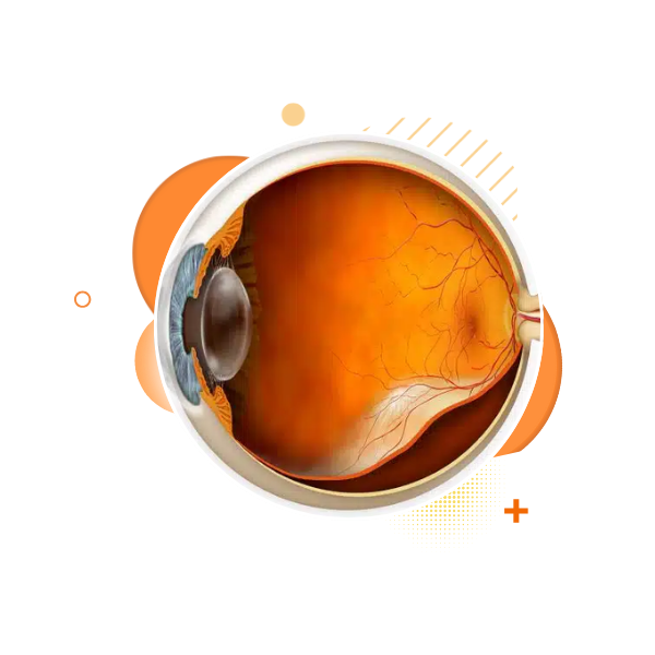
Retinal Detachment
What Is Retinal Detachment?
Retinal detachment is a serious eye condition in which the retina separates from its normal position at the back of the eye.
The retina is the light-sensitive layer that sends visual signals to the brain. When it detaches, it loses its blood supply and oxygen, causing sudden vision problems.
⚠️ Retinal detachment is a medical emergency. If not treated promptly, it can lead to permanent vision loss or blindness.
» Why Is the Retina So Important?
The retina works like a camera sensor:
It captures light entering the eye
Converts it into signals
Sends these signals to the brain for vision
When the retina detaches, it cannot function properly, leading to partial or complete loss of sight.
» Common Causes of Retinal Detachment
Retinal detachment can occur due to several reasons, including:
Aging changes in the eye
High myopia (high minus power)
Eye injury or trauma
Previous eye surgery (e.g., cataract surgery)
Diabetic eye disease
Weak areas or tears in the retina
Family history of retinal detachment
The risk increases with age and certain eye conditions.
» Types of Retinal Detachment
‣ Rhegmatogenous Retinal Detachment
Most common type
Occurs due to a tear or hole in the retina
Fluid enters through the tear and lifts the retina
‣ Tractional Retinal Detachment
Caused by scar tissue pulling on the retina
Common in advanced diabetic retinopathy
‣ Exudative (Serous) Retinal Detachment
Caused by fluid leakage under the retina
No tear or hole present
Seen in inflammation, tumors, or severe BP problems
» Warning Signs & Symptoms
Retinal detachment often begins suddenly. Seek urgent eye care if you notice:
Sudden increase in floaters (black spots or cobwebs)
Flashes of light, especially in side vision
A dark shadow or curtain coming over vision
Sudden blurred or reduced vision
Loss of side (peripheral) vision
⚠️ Pain is usually absent, so do not ignore visual symptoms.
» Who Is at Higher Risk?
You may be at higher risk if you:
Have high minus power (myopia)
Have diabetes
Had eye injury or surgery
Have retinal thinning or weak retina
Have family history of retinal detachment
Are above 50 years of age
Regular eye check-ups are important for high-risk individuals.
» How Is Retinal Detachment Diagnosed?
Diagnosis is done through a detailed dilated retinal examination.
Additional tests may include:
Retinal imaging
Optical Coherence Tomography (OCT)
Ultrasound of the eye (if media is cloudy)
Early diagnosis greatly improves the chances of saving vision.
» Management & Care (Overview)
Retinal detachment does not improve on its own.
Management depends on the type, size, and duration of detachment.
General goals of care:
Reattach the retina
Prevent further retinal damage
Preserve as much vision as possible
Early intervention leads to better visual outcomes.
» Recovery & Follow-Up
Vision recovery varies from patient to patient
Some blurring may persist initially
Regular follow-ups are essential
Protecting the eye and following medical advice is crucial
Vision outcome depends on how early the retina was treated and whether the central retina (macula) was involved.
» When Should You See an Eye Doctor Immediately?
Seek urgent care if you experience:
Sudden flashes or floaters
Shadow or curtain over vision
Sudden vision loss in one eye
🚨 Do not wait—early action can save eyesight.
Retinal Detachment FAQs
Retinal detachment is a condition where the retina becomes separated from the underlying tissue, leading to potential vision loss. It is a medical emergency that requires prompt treatment to avoid permanent damage to vision.
Symptoms include flashes of light, the sudden appearance of floaters, blurred or distorted vision, a curtain or shadow in the vision, and loss of peripheral vision.
Retinal detachment is diagnosed through a comprehensive eye exam, including a dilated eye exam, optical coherence tomography (OCT), ultrasound, and sometimes fundus photography.
Retinal detachment can be caused by aging, eye injury, severe nearsightedness (myopia), or underlying health conditions such as diabetes. It can also occur due to the formation of retinal tears or holes.
Treatment for retinal detachment includes laser surgery, cryotherapy, pneumatic retinopexy, scleral buckling, or vitrectomy, depending on the severity and type of detachment.